A basic blood test that evaluates red cells, hemoglobin, white cells and platelets. It can point to anemia, inflammation, infection effects, blood loss and bone-marrow stress, but it does not explain the cause by itself.
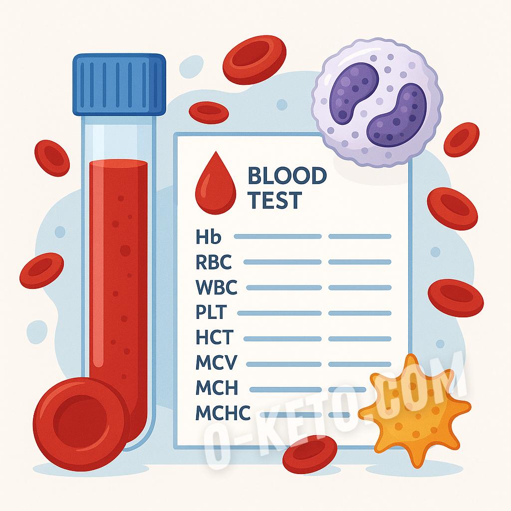
A complete blood count evaluates the cellular part of blood: red blood cells, hemoglobin, hematocrit, red-cell indices, white blood cells with differential and platelets. It is not a test for everything, but a quick way to see how the body is handling inflammation, deficiencies, blood loss, bone-marrow load and some medication effects.
The common mistake is to look only at numbers flagged in red. The report has to be read as a system: hemoglobin without MCV and RDW says little about anemia type, white cells without a differential do not describe the reaction, and platelets without symptoms do not explain bleeding or clotting risk.
Red-cell part
Hemoglobin shows the oxygen-carrying capacity of blood, hematocrit reflects the cellular share of blood volume, and the red-cell count helps judge the degree of change. MCV describes average red-cell size: small cells often raise questions about iron deficiency or chronic inflammation; large cells point more toward B12, folate, alcohol, liver disease, hypothyroidism or medication effects.
MCH and MCHC describe hemoglobin content in red cells, while RDW shows how varied cell size is. A high RDW with still-normal hemoglobin may appear early in iron deficiency or with mixed deficiencies. Therefore weakness, shortness of breath, hair loss, brittle nails and palpitations may deserve more than “hemoglobin is normal”.
White cells and differential
The white-cell differential shows proportions of neutrophils, lymphocytes, monocytes, eosinophils and basophils. Neutrophils often rise with bacterial infection, acute inflammation, stress and glucocorticoids. Lymphocytes may increase with viral infections and some immune states, and decrease after severe stress, illness or medication load.
Eosinophils matter in allergy, parasitic infection and some inflammatory diseases. Monocytes often shift during recovery after infection. But the differential does not replace clinical assessment: the same number can mean different things in someone with fever, in an athlete after hard training and in a patient taking medication.
Platelets and clotting context
Platelets help stop bleeding, but a complete blood count is not a full coagulation panel. Low platelets matter with bruising, gum bleeding, nosebleeds, heavy periods and before procedures. High platelets may be a reaction to inflammation, iron deficiency, recent blood loss or, less commonly, a bone-marrow disorder.
If platelets are very different from previous values, the test is often repeated because laboratory artifacts are possible: cell clumping in the tube, delayed processing or anticoagulant effects. When the change is real, clinicians usually compare the CBC with ferritin, CRP, coagulation tests, liver markers and medication history.
Preparation and distortions
Acute infections, poor sleep, hard training the day before, dehydration, blood loss, menstruation, pregnancy, smoking, alcohol, iron, B12, folate, hormones, anticoagulants and anti-inflammatory drugs can change the result. After illness, values may remain shifted for days or weeks, so a single report without context is easy to overread.
For a routine check, blood is best drawn in the morning, in a calm state, without heavy training the day before and without trying to “improve” the test with water, supplements or fasting. If the test is ordered during an acute condition, preparation is secondary because the real illness picture is the point.
Low-carb context
Keto or LCHF should not by themselves cause anemia or major white-cell shifts. But if the diet becomes poor in vegetables, organ meats, fish and complete protein, CBC changes may reveal iron, B12 or folate problems. This is especially relevant when someone removes meat, liver, eggs or dairy and leaves only fat, coffee and a narrow set of foods.
Weakness, shortness of breath, palpitations, unusual bruising, prolonged fever, night sweats, enlarged lymph nodes or unexplained weight loss need medical interpretation. Taking iron, B12 or folate based on one number can hide blood loss, inflammation, thyroid disease, malabsorption or another cause.
What to compare it with
If the CBC suggests anemia, ferritin, serum iron, transferrin or TIBC, B12, folate and inflammation markers are often needed. With white-cell changes, symptoms, temperature, CRP, ESR, recent infections and medication history matter. With platelet changes, coagulation tests, liver markers, ferritin and bleeding history help. This does not make the test unnecessarily complicated; it prevents the main mistake, which is treating a number without understanding why it moved.
Published: 26.10.2025
Updated: 07.06.2026
Any remaining questions? Ask chatGPT.:
If you have any questions about the term "Complete blood count", you can ask them to AI. Please note, a low-cost OpenAI model is used. It may answer questions about disease treatment with errors!
Increased hematocrit may reflect relative blood concentration from insufficient fluid intake; interpret it together with hemoglobin, erythrocytes, hydration, and overall condition.
Platelets above the upper limit may be a reactive shift in hidden or overt iron deficiency; compare the result with ferritin, hemoglobin, MCV, and inflammatory context.
Increased mean hemoglobin content in erythrocytes may accompany vitamin B12 deficiency; compare it with MCV, hemoglobin, homocysteine, and folate status.
Reduced erythrocytes may accompany vitamin B12 deficiency, especially when MCV is increased or there are signs of megaloblastic hematopoiesis; compare with B12, folic acid, and homocysteine.
Reduced erythrocytes may accompany folic acid deficiency, especially with macrocytic shifts; evaluate together with B12, homocysteine, and erythrocyte indices.
Increased MPV together with macrocytic changes may accompany vitamin B12 deficiency and requires assessment of B12, folic acid, homocysteine, and the overall CBC pattern.
Reduced platelets may accompany vitamin B12 deficiency, especially when macrocytosis, low erythrocytes, or other signs of B vitamin deficiency are present.
Reduced hematocrit may accompany anemia, especially when hemoglobin or erythrocytes are also reduced; the cause should be clarified with ferritin, B12, folate, and erythrocyte indices.
Increased hematocrit may accompany polycythemia when the bone marrow produces too many erythrocytes; compare with hemoglobin, erythrocytes, hydration, and thrombosis risk.
Increased platelets after a viral infection may reflect post-viral recovery and endothelial response; compare with symptoms, leukocyte formula, and coagulation markers.
Increased erythrocytes may accompany polycythemia and requires assessment of hematocrit, hemoglobin, symptoms of blood viscosity, and causes of secondary erythrocytosis.
This macrocytic profile may accompany megaloblastic anemia and requires assessment of vitamin B12, folic acid, homocysteine, and other erythrocyte indices.
Increased erythrocytes may reflect relative blood concentration with dehydration; interpret together with hematocrit, hemoglobin, platelets, and fluid intake.
This hemoglobin level may fit iron deficiency anemia when it appears together with depleted iron stores: low ferritin, microcytosis, reduced MCH, or low transferrin saturation.
Low erythrocytes can be part of the iron deficiency anemia pattern and should be interpreted together with hemoglobin, hematocrit, MCV, MCH, and ferritin.
Reduced mean corpuscular hemoglobin supports a hypochromic iron deficiency pattern and is interpreted together with MCV, ferritin, and transferrin saturation.
Low reticulocyte hemoglobin indicates insufficient iron delivery to newly formed erythrocytes and may reveal iron deficient erythropoiesis before major hemoglobin changes.
This platelet level does not confirm thrombosis by itself, but it may accompany higher thrombotic risk and requires assessment of thrombocytosis causes, inflammation, iron status, and coagulation markers.
Elevated basophils in a complete blood count may accompany allergic and histamine-driven reactivity; the result should be assessed together with eosinophils, IgE, skin, nasal and bronchial symptoms, and medication use.
Hypersegmented neutrophils on a blood smear may support suspected vitamin B12 deficiency and a megaloblastic hematopoietic pattern; assess it together with MCV, MCH, white cells, platelets, B12, folate, and clinical context.
Hypersegmented neutrophils may occur with folate deficiency and megaloblastic hematopoiesis; compare the result with MCV, MCH, RDW, platelets, folate level, B12, and causes of impaired absorption.
Acute stress before blood draw
Neutrophils
This pattern may reflect a cortisol-driven stress response before the blood draw; compare it with symptoms, physical exertion, sampling time, and a repeat CBC under calmer conditions.
During pregnancy, a moderate decrease in hemoglobin may reflect physiologic hemodilution; it should be distinguished from true iron deficiency using ferritin, red cell indices, and the overall clinical context.
This level corresponds to severe anemia and requires urgent medical assessment of blood loss, deficiencies, hemolysis, or other disorders of blood formation.
Hematocrit below 40% may accompany anemia or blood loss; interpret it together with hemoglobin, erythrocytes, MCV, MCH, and the clinical context.
Lab test interpretations are provided for informational purposes only. Do not interpret results on your own if you do not clearly understand what you are doing, are not a nutrition professional, or have not consulted a physician.
List of videos from YouTube by request "Complete blood count":




 1 / 4
1 / 4


 1 / 4
1 / 4